 Understanding the Process of Corneal Healing
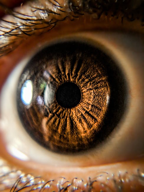
Just How to avoid Corneal Scarring When it’s healthy, the cornea is a clear, dome-shaped tissue that secures your eyes from dirt and also various other particles. But when it’s harmed, your vision can end up being blurry. This kind of scar can be brought on by abrasions, lacerations, or burns. Your ophthalmologist can examine your cornea for marks making use of a slit lamp and also special dyes. These examinations can reveal any changes in the shape of your cornea or opacity of the front surface area. Prevention is always the easiest way to prevent corneal scarring, yet it’s additionally essential to capture as well as deal with any eye injuries, infections or various other issues that might create the cornea to end up being harmed. Therapies include antibiotics, antiviral eye drops as well as steroid eye decreases, which can minimize the likelihood of scarring. The cornea has a complex structure that includes a central round tissue called the epithelium and also surrounding layers that create a barrier to secure the eye from infection. These layers are made of a layer of cells called the keratocytes, which have highly revealed healthy proteins known as crystallins that contribute to the transparent nature of the cornea. During a slit lamp exam, your optometrist will look for a white, nontransparent line that suggests a mark on your cornea. They’ll also look for any kind of other adjustments in the surface area of your cornea, such as modifications in shade or the appearance of a bump. A corneal scar can intensify with time, specifically if it’s connected with a chronic problem like keratoconus. In these situations, it’s important to see your eye doctor as soon as possible to make sure that your medical professional can figure out the cause of the scar and also establish the best treatment plan for your eyes. As a component of injury recovery, the corneal surface area undertakes an intricate series of occasions that includes removal of the hurt cells, cell proliferation and migration to the injury website, and also repair service and replacement before the injury heals. During this procedure, a variety of cell kinds are entailed that lead to the formation of new collagen fibers that strengthen the cornea as well as restore its look. Collagen is the most common sort of tissue in your eye and it comprises regarding 90 percent of the cornea’s external surface area. This tissue can be thick or slim and it is composed of parallel bundles of fibers (fibroils) that are secreted by the keratocytes. These fibers are crucial for the bending of light and also concentrating pictures on the retina in the back of your eye. When the keratocytes are damaged, they don’t create enough of these fibers to correct the issue. This is why the scars that develop over your cornea are commonly thicker than typical. It is necessary to get a full eye test every year to stop corneal damages and also maintain your eyes healthy and balanced. The ophthalmologist will certainly utilize the slit lamp and also an unique tool called an ophthalmoscope to check for any type of changes in your eye’s framework or other indications of illness.